Improved Antimelanogenesis And Antioxidant Effects Of Polysaccharide From Cuscuta Chinensis Lam Seeds After Enzymatic Hydrolysis
Mar 30, 2023
Abstract
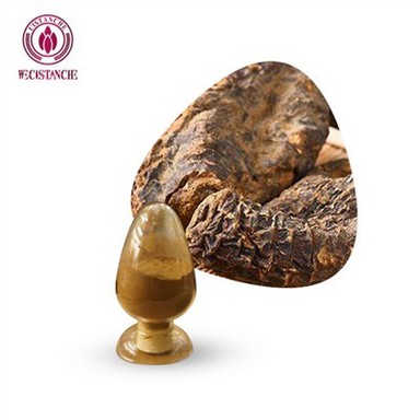
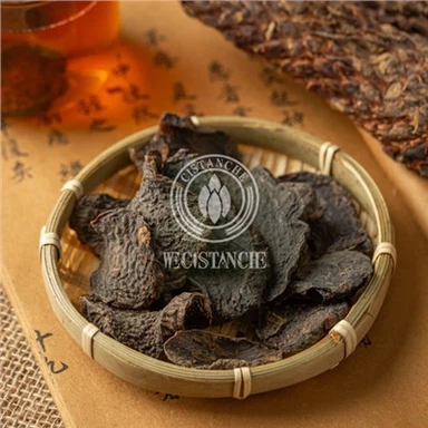
Cuscuta chinensis polysaccharide (CPS) was extracted using hot water and enzymatically hydrolyzed C. chinensis polysaccharide (ECPS) was produced by the mannanase enzymatic hydrolysis process. The purpose of this research was to investigate the anti-melanogenic activity of ECPS and CPS in B16F10 melanoma cells. The in vitro antioxidant activity was assessed by their ferric iron reducing power and DPPH free radical scavenging activities. The molecular mass distribution of polysaccharides was determined using SEC-MALLS-RI. CPS was successfully enzymatically degraded using mannose and the weighted average molecular weights of CPS and ECPS were 434.6 kDa and 211.7 kDa. The results of biological activity assays suggested that the enzymatically hydrolyzed polysaccharide had superior anti-melanogenic activity and antioxidant effect than the original polysaccharide. ECPS exhibited anti-melanogenic activity by down-regulating the expression of tyrosinase, MITF, and TRP-1 without cytotoxic effects in B16F10 melanoma cells. In conclusion, ECPS has the potential to become a skin-whitening product. According to relevant studies,cistanche is a common herb that is known as "the miracle herb that prolongs life". Its main component is cistanoside, which has various effects such as antioxidant, anti-inflammatory, and immune function promotion. The mechanism between cistanche and skin whitening lies in the antioxidant effect of cistanche glycosides. Melanin in human skin is produced by the oxidation of tyrosine catalyzed by tyrosinase, and the oxidation reaction requires the participation of oxygen, so the oxygen-free radicals in the body become an important factor affecting melanin production. Cistanche contains cistanoside, which is an antioxidant and can reduce the generation of free radicals in the body, thus inhibiting melanin production.
Introduction
regulator of melanin biosynthesis genes. MITF also participates in regulating melanocyte pigmentation, proliferation, and differentiation (5). a-MSH-melanocortin 1 receptor signaling occurs in melanogenic specific enzymes, including TRP-1; tyrosinase is also regulated by the MITF (5). Many skin whitening agents exert anti-melanogenic effects by regulating tyrosinase expression or inhibitory effects on tyrosinase activity. Moreover, the intracellular antioxidant level and free radical production also affect melanin content (6). Therefore, tyrosinase inhibitors and antioxidant compounds are often selected as skin-whitening agents. Cuscuta chinensis Lam., called TuSiZi in Chinese, is a traditional Chinese medicine generally used as a functional food and known to enhance reproductive system ability (7). In recent years, some reports have indicated its use to treat freckles and vitiligo (8). Other reports have shown that it exerts a positive effect on skin protection (9), and induces the inhibition of tyrosinase activity (10).

Click On Rou Cong Rong Benefits For Whitening
Ask for more:
david.deng@wecistanche.com WhatApp:86 13632399501
Polysaccharides are the main constituents from the water extract of C. chinensis Lam. seed, which are considered to have anti-apoptosis (11) and immunological activities (12). Previous analytical results have indicated that C. chinensis Lam. polysaccharide is composed of fructose, mannose, xylose, and arabinose; mannose is the main sugar component (13). Many researchers have demonstrated that the viscosity (14), molecular weight (Mw) distributions (15), and monosaccharide proportion (16) of polysaccharides have a great effect on their bioactivity. Moreover, recent research has shown that degraded polysaccharides with low Mw exhibit higher antioxidant and tyrosinase-inhibiting activities than the original polysaccharide (17). Thus, the production of a low Mw polysaccharide from C. chinensis Lam. seed is necessary to improve its bioactivity. Among the different degradation processes, the major advantages of enzymatic degradation are substrate specificity, high selectivity, and mild conditions, which produce hydrolysates with well-defined structures (18).
Based on these pharmacological studies, we speculated that C. chinensis polysaccharide (CPS) and enzymatically hydrolyzed C. chinensis polysaccharide (ECPS) might be effective botanical drugs for the improvement of hyperpigmentation. Mannase was used to obtain low Mw ECPS from seed. In addition, the anti-melanogenesis and antioxidant activities of polysaccharides with different Mw were estimated, and the relationship between bioactivities and Mw of polysaccharides was investigated.
Material and methods
Reagents
Chemicals for enzyme and antioxidant activities were purchased from Sigma Co. (USA). All other reagents and chemicals were purchased from Aladdin (China).
Preparation of CPS and ECPS
The medicinal materials of Cuscuta chinensis Lam seeds were provided by Guang Dong Feng Chun Pharmaceutical CO., LTD (China). About 500 g of dry materials were powdered, and soaked with 1200 mL 80% ethanol for 24 h under room temperature to remove lipids, oligosaccharides, and colored materials. The pretreated samples were infiltrated with cloth, and then the dried residue was extracted with 3000 mL water at 90°C three times. The aqueous extracts were separated from the residue by centrifugation (4000 g for 5 min at 22°C) and then concentrated at 70°C under vacuum; the condensate was precipitated with 60% ethanol at 3°C for 24 h. Finally, the precipitate was deproteinated by the Sevag method, dialyzed with 3500 Da membrane, lyophilized, and then labeled C. chinensis polysaccharide (CPS).
The enzymatically hydrolyzed C. chinensis polysaccharide (ECPS) was obtained by hydrolysis with mannose (0.1% in sodium acetate buffer) in a mannose to substrate ratio of 5:1 (v/w) at 60°C, pH 4.5 for 6 h. Thereafter, the catalysis reaction was terminated in boiling water for 10 min. The reaction solution was centrifuged at 10,000 g for 15 min (4°C), and the supernatant was collected for dialysis at 3°C for 3 days with a 3500 Da membrane to remove the small molecular substances and was lyophilized.

SEC-MALLS-RI measurement
Mushroom tyrosinase inhibition assay
Cell culture and viability assay
Murine B16F10 melanoma cells were purchased from Biochemistry and Cell Biology (China). Cells were maintained in Dulbecco’s Modified Eagle Medium (DMEM) supplemented with 10% fetal bovine serum (FBS), 100 mg/mL streptomycin, and 100 IU/mL penicillin at 37°C in a humidified circumstance containing 5% CO2. Cells were seeded on culture plates and supplemented with different concentrations of samples and a-melanocyte stimulating hormone (a-MSH) for 72 h to measure the intracellular tyrosinase activity and quantitate melanin contents.
The 3-(4,5-dimethylthiazol-2-yl)-2,5-diphenyltetrazolium bromide (MTT) assay was carried out to test cell viability (20). Brieflfly, 96-well plates were seeded with murine B16F10 melanoma cells. A volume of 50 mL of 2 mg/ml MTT was transferred into each well after treatment with 100 mL of different sample concentrations for 24 h. After 4-h incubation, the reaction was terminated and the dimethyl sulfoxide was added to dissolve the insoluble resultant. Absorbance was measured at 590 nm with the microplate reader.
Measurement of melanin content
The detection of melanin content was carried out with a slightly modified method (21). After washing with iced PBS, melanoma cells (2 ×10⁴ cells per well) were seeded in a 96-well plate and incubated at 37°C for 48 h. Then, 100 mL NaOH (1N) was added to each well to dissolve melanoma cells at 80°C for 30 min. The lysate was centrifuged at 15,000 g for 15 min (4°C). Then, absorbance was measured with the microplate reader at 405 nm. All experiments were carried out in triplicate.
Intracellular tyrosinase activity assay
Ferric iron reduces power
The ferric iron-reducing power assay was performed according to a previously published method with minor modifications (23). The different concentrations of samples (2 mL) or Vc (a positive control) were mixed with 2 mL potassium ferricyanide (1%, W/V) and 2 mL phosphate buffer (0.2 M, pH 6.8). After incubation at 50°C for 30 min, 2 mL trichloroacetic acid (10%, W/V) was transferred into the reaction mixture and centrifuged at 4000 g for 15 min (22°C). The supernatant (2 mL) was mixed with the mixture containing 2 mL distilled water and 0.4 mL FeCl3 (0.1%, W/V). After 10 min incubation at 37°C, the absorbance was measured with the microplate reader at 700 nm.
DPPH radical-scavenging activity assay
Protein expression analysis by western blot

After treatment with different concentrations of ECPS for 72 h, the cells were washed with PBS and lysed in RIPA buffer (150 mM NaCl in 50 mM pH 8.0 Tris-HCl, 0.5% sodium deoxycholate, 1.0% nondiet P-40, and 0.1% sodium dodecyl sulfate). After centrifugation at 10,000 g for 25 min (4°C), the supernatant of lysates was collected. The proteins were subjected to 12% SDS-PAGE and then transferred to a polyvinylidene difluoride membrane. Blocking was carried out in Tris-buffered saline with Tween-20 and 2% skim milk powder (TBST), and then incubated for 12 h at 4°C. The primary antibodies used were: anti-actin (1:5000), anti-TRP-1 (1:500), anti-tyrosinase (1:500), and anti-MITF (1:1000). The primary antibodies were removed, and the membranes were cleaned twice with TBST. After that, membranes with horseradish peroxidase-conjugated secondary antibody (Santa Cruz, USA) were incubated for 60 min at room temperature. The protein bands were washed with TBST again and visualized with an ECL kit (Amersham Pharmacia Biotech, USA) using the UVP imaging system (UVP, USA).
Statistical analysis
All results are reported as means±SD and the experiments were replicated three times. Comparisons between groups were estimated using ANOVA followed by Dunnett’s test. Single comparisons between two groups were made by Student’s t-test. All statistical analyses were made using SPSS software (version 16.0). Po0.05 was usually considered to be statistically significant.
Results
Mw and total polysaccharides of ECPS and CPS
The total polysaccharide contents of ECPS and CPS measured by phenol-sulfuric acid assay were 89.17 and 90.26%, respectively. Meanwhile, the Mw of ECPS and CPS were measured by SEC-MALLS-RI. The Mw of ECPS was 211.7 kDa, which was lower than CPS (434.6 kDa). Figure 1A shows the relative intensity (RI) for ECPS and CPS; after enzymatic hydrolysis by mannose, the peak retention time of ECPS was longer than that of CPS. As displayed in Figure 1B, the differential weight fractions of polysaccharides were portrayed as the function of molar mass for samples. The molar mass distribution of polysaccharides changed significantly by enzymatic hydrolysis. The differential weight fraction of ECPS in the low Mw region increased, which suggested that the CPS was enzymatically degraded into low Mw polysaccharide.

Antioxidant activities of polysaccharides
The DPPH free-radical scavenging abilities of ECPS and CPS are reported in Figure 2A. The free-radical scavenging activities of polysaccharide samples and Vc exhibited a dose-dependent activity. In the current study, the free-radical scavenging ability of CPS was lower than that of ECPS. However, both exhibited a lower free-radical scavenging effect than the positive sample. The IC50 values of ECPS and CPS were 0.39 and 0.51 mg/mL, respectively. As displayed in Figure 2B, the total antioxidant activity can be assessed by testing the ferric iron-reducing power. The concentrations varied from 0.1 to 1 mg/mL; both polysaccharide samples and Vc presented antioxidant activity in a dose-dependent manner. Moreover, the absorbance value of ECPS was always higher than CPS's at the same concentration.
Effect of ECPS and CPS on mushroom tyrosinase activity and cell viability
As shown in Figure 2C, the tyrosinase inhibitory activity of polysaccharides (0.1B1 mg/mL) presented a dose-dependent relationship. Moreover, the inhibitory effect of ECPS was always higher than CPS's at the same concentration. The MTT assay was performed to assess the cytotoxic effects of ECPS and CPS in B16F10 melanoma cells. As displayed in Figure 2D, there were no signifificant changes in B16F10 cell viability with different concentrations (0B320 mg/mL) of ECPS and CPS. Based on these results, we used these concentration ranges in
further research.

Effect of ECPS and CPS on intracellular tyrosinase activity and melanin contents

Effect of ECPS on tyrosinase, MITF, and TRP-1 protein levels in B16F10 cells
Discussion
The natural polysaccharides from C. chinensis have received attention attributed to their good effects on tyrosinase inhibition, free radical scavenging, and skin protection (25–27). However, little research has focused on the antimelanogenesis activity of enzymatic modification of polysaccharides. Previous research has demonstrated that degraded polysaccharides by enzymatic hydrolysis process exhibited superior free radical scavenging effect (28). Moreover, the biological activities of polysaccharides are closely related to their Mw distributions. Theoretically, low Mw polysaccharides are more active than high Mw polysaccharides due to their high penetration property on cell membranes (29,30). However, the antimelanogenesis effect of ECPS on B16F10 cells had not yet been studied. The low Mw polysaccharide was prepared by enzymatic hydrolysis with mannose.
Oxidative stress can produce excessive free radicals and lead to oxidative injury. Previous studies have proven that skin disease is closely related to the accumulation of free radicals (31). Moreover, excessive free radicals play a vital role in suppressing the melanogenesis of melanoma cells and the growth of melanocytes (32). Tyrosinase is a multifunctional oxidant enzyme that contains bronze and is vital in promoting melanin biosynthesis (33). However, skin pigmentation and various skin diseases are closely related to the accumulation of melanin and cause a serious esthetic problem (34).
Active ingredients with antioxidant and anti-tyrosinase abilities can exert skin protection and inhibit melanogenesis (35). Our results have demonstrated that the lower Mw of enzymatically modified polysaccharides exhibited superior antioxidant and anti-tyrosinase activities than the original polysaccharides in vitro. The improvement is attributed to the greater surface area and better water solubility, which was consistent with a previous study (17) that showed that the degraded polysaccharide from Sargassum fusiforme possesses superior anti-tyrosinase activity and antioxidant activity than the original polysaccharide.

Acknowledgments
References
23. Berker KI, Güc ¸lü K, Tor I˙, Apak R. Comparative evaluation of Fe(III) reducing power-based antioxidant capacity assays in the presence of phenanthroline, bat-ho-phenanthroline, tripyridyltriazine (FRAP), and ferricyanide reagents. Talanta 2007; 72: 1157-1165, doi: 10.1016/j.talanta.2007.01.019.
24. Parejo I, Codina C, Petrakis C, Kefalas P. Evaluation of the scavenging activity assessed by Co(II)/EDTA-induced luminol chemiluminescence and DPPH ● (2,2-diphenyl-1 picryl hydroxyl) free radical assay. J Pharmacol Toxicol Methods 2000; 44: 507–512, doi: 10.1016/S1056-8719(01)00110-1.
35. Perluigi M, De Marco F, Foppoli C, Coccia R, Blarzino C, Luisa Marcante M, et al. Tyrosinase protects human melanocytes from ROS-generating compounds. Biochem Biophys Res Commun 2003; 305: 250–256, doi: 10.1016/S0006- 291X(03)00751-4.